ᱨᱮᱫ:3D CT of thorax, annotated.jpg

ᱧᱮᱞᱡᱚᱝ ᱨᱮᱱᱟᱜ ᱟᱠᱟᱨ:᱘᱐᱐ × ᱕᱘᱖ ᱯᱤᱠᱥᱮᱞ ᱮᱴᱟᱜ ᱨᱤᱡᱚᱞᱩᱥᱚᱱᱥ: ᱓᱒᱐ × ᱒᱓᱔ ᱯᱤᱠᱥᱮᱞ | ᱖᱔᱐ × ᱔᱖᱙ ᱯᱤᱠᱥᱮᱞ | ᱑,᱐᱒᱔ × ᱗᱕᱐ ᱯᱤᱠᱥᱮᱞ | ᱑,᱒᱘᱐ × ᱙᱓᱘ ᱯᱤᱠᱥᱮᱞ | ᱑,᱕᱔᱔ × ᱑,᱑᱓᱑ ᱯᱤᱠᱥᱮᱞ
ᱟᱥᱚᱞ ᱨᱮᱫ (᱑,᱕᱔᱔ x ᱑,᱑᱓᱑ pixels, file size: ᱖᱕᱖ KB, MIME type: image/jpeg)
ᱢᱩᱬᱩᱛ ᱠᱟᱛᱷᱟ
| ᱵᱤᱵᱚᱨᱚᱱᱤ |
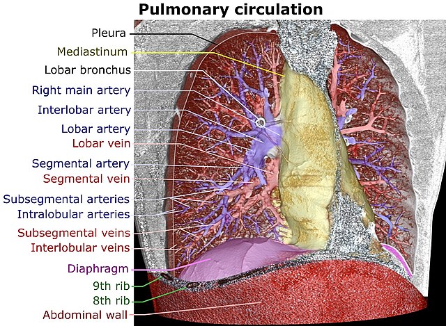
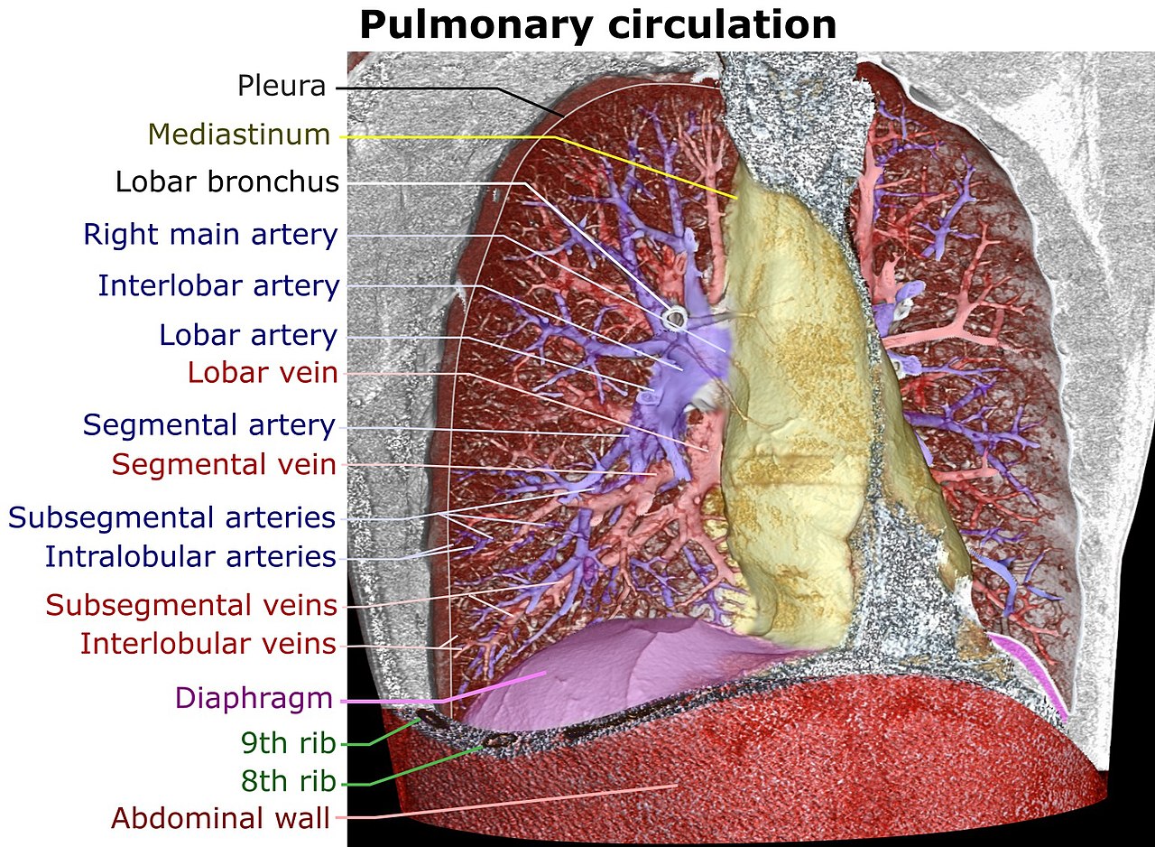
English:
|
| ᱢᱟᱹᱦᱤᱛ | |
| ᱯᱷᱮᱰᱟᱛ |
ᱤᱧᱟᱜ ᱠᱟᱹᱢᱤ
|
| ᱚᱱᱚᱞᱤᱭᱟᱹ | Mikael Häggström |
| Other versions |
|


ᱞᱟᱭᱥᱮᱱᱥ ᱛᱮᱭᱟᱨ
I, the copyright holder of this work, hereby publish it under the following license:
| This file is made available under the Creative Commons CC0 1.0 Universal Public Domain Dedication. | |
| The person who associated a work with this deed has dedicated the work to the public domain by waiving all of their rights to the work worldwide under copyright law, including all related and neighboring rights, to the extent allowed by law. You can copy, modify, distribute and perform the work, even for commercial purposes, all without asking permission.
|
ᱨᱮᱫ ᱨᱮᱭᱟᱜ ᱱᱟᱜᱟᱢ
ᱚᱠᱛᱚ ᱨᱮ ᱞᱤᱱ ᱢᱮ/ᱚᱠᱛᱚ ᱨᱮ ᱨᱮᱫ ᱧᱮᱞ ᱞᱟᱹᱜᱤᱛ ᱞᱤᱱ ᱢᱮ
| ᱢᱟᱹᱦᱤᱛ/ᱚᱠᱛᱚ | ᱴᱤᱯ | ᱡᱚᱠᱷᱟ | ᱵᱮᱵᱷᱟᱨᱤᱭᱟᱹ | ᱠᱟᱛᱷᱟ | |
|---|---|---|---|---|---|
| ᱱᱤᱛᱚᱜ | ᱐᱖:᱒᱘, ᱒᱕ ᱡᱩᱱ ᱒᱐᱑᱗ | | ᱑,᱕᱔᱔ × ᱑,᱑᱓᱑ (᱖᱕᱖ KB) | Mikael Häggström | + Lobular |
| ᱑᱙:᱓᱔, ᱒᱔ ᱡᱩᱱ ᱒᱐᱑᱗ |  | ᱑,᱖᱔᱔ × ᱑,᱐᱖᱒ (᱖᱓᱒ KB) | Mikael Häggström | Corrected - there is very seldom a main pulmonary vein. Also, there's an interlobar artery segment on the right lung | |
| ᱑᱔:᱓᱗, ᱒᱔ ᱡᱩᱱ ᱒᱐᱑᱗ |  | ᱑,᱖᱔᱔ × ᱑,᱐᱖᱒ (᱖᱓᱙ KB) | Mikael Häggström | User created page with UploadWizard |
ᱯᱷᱟᱭᱤᱞ ᱵᱮᱣᱦᱟᱨ
ᱞᱟᱛᱟᱨ ᱨᱮᱭᱟᱜ ᱥᱟᱦᱴᱟ ᱡᱚᱱᱚᱲᱠᱚ ᱱᱤᱭᱟᱹ ᱨᱮᱫ ᱨᱮ:
ᱡᱮᱜᱮᱛ ᱡᱟᱠᱟᱛ ᱨᱮᱫ ᱵᱮᱵᱷᱟᱨᱟᱜ
ᱱᱚᱶᱟ ᱨᱮᱫᱠᱚ ᱵᱮᱵᱷᱟᱨᱟᱠᱟᱫ ᱣᱤᱠᱤᱠᱚ :
- ar.wikipedia.org ᱨᱮ ᱵᱮᱣᱦᱟᱨ
- bn.wikipedia.org ᱨᱮ ᱵᱮᱣᱦᱟᱨ
- en.wikipedia.org ᱨᱮ ᱵᱮᱣᱦᱟᱨ
- it.wikipedia.org ᱨᱮ ᱵᱮᱣᱦᱟᱨ
- ko.wikipedia.org ᱨᱮ ᱵᱮᱣᱦᱟᱨ
- ml.wikipedia.org ᱨᱮ ᱵᱮᱣᱦᱟᱨ
- ro.wikipedia.org ᱨᱮ ᱵᱮᱣᱦᱟᱨ
- vi.wikipedia.org ᱨᱮ ᱵᱮᱣᱦᱟᱨ